3D Localization of Single Molecules Improved to Nanoscale

University researchers W. E. Moerner, E. Betzig and S. W. Hell won the 2014 Nobel Prize for their development of super-resolution microscopy, a technique that breaks the optical diffraction limit following fluorescent emissions from single molecules. Now, the team led by Professor Moerner at Stanford University has improved this technology, achieving unprecedented accuracy when tracking single molecules in 3D. The study has been published in the journal Optica.
Tracking single molecules discovers very valuable information about cell signaling, neuron communication molecular structure and dynamics. In the last years, single molecule localization has vastly improved thanks to super-resolution microscopy. In this technique, single molecules are stimulated to fluoresce asynchronically, avoiding the posible confusion caused by overlapping signals from close molecules. With this method, sub-diffraction resolution imaging is obtained. With the right function, the data can inform even about 3D structure. However, this information is not very accurate, due to suboptimal calibration techniques.
Improving calibration techniques
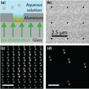
 With current calibration techniques (fluorescent beads), single molecule signals can have a localization precision of 10 nm. This can lead to wrong interpretations about the number of molecules and their dynamics, especially in 3D. In their last study, Woerner and colleagues developed a new microscope calibration technique that would correct optical distortion across the whole field of view. Instead of beads, they used a metallic plate with an array of nanoholes filled up with fluorescent dye. The new system reduced distortion, allowing the best 3D single molecule localization ever. The new calibration also reduced the error when using point spread functions for 3D localization.
In the picture, a nanohole is represented (a). The light, in green, passes through the coverslip and reaches the flourescent dye contained in the hole practiced in an aluminum layer. The points of light (orange) are detected from below. (b) is a scanning electron microscope image of the nanoholes array (each hole is 200 nm in diameter).
The researchers are currently using the new calibration method in all their single molecule and super-resolution experiments, like a study of single molecule dynamics in a 2 micron bacteria.
Source: OSA

