A smartphone accessory to detect sickle cell disease


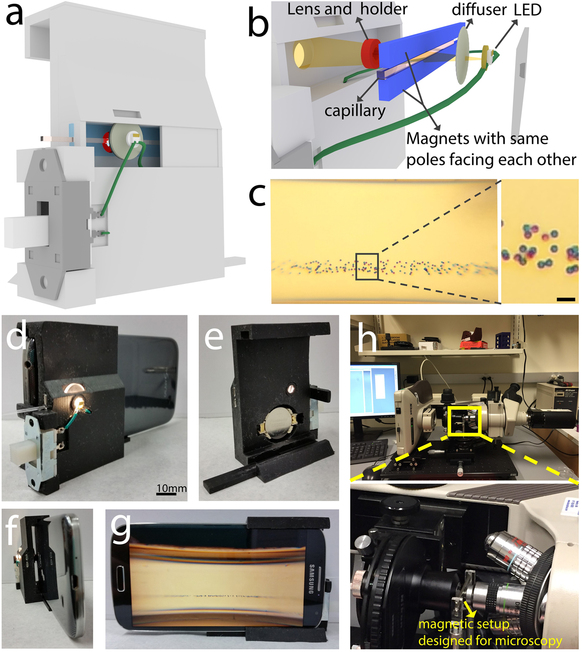
Design and images of the Sickle Cell Detector (a,b, d-g); polystyrene microspheres levitating (c); magnetic setup in a microsope (h). Credit: Scientific Reports.
A team of scientists from four top American universities have developed a smartphone attachment that allows to detect sickle cell disease. The 3D printed device uses the phone´s camera and an application. Composed of magnets, a microcapillary and optical components, it can detect the density of red blood cells in deoxygenated conditions and thus identify the denser sickle cells. The results have been published in the journal Scientific Reports.
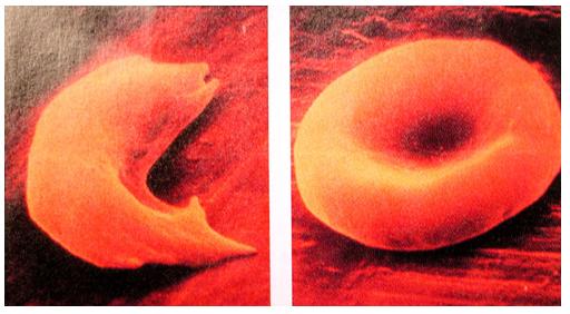
Sickle cell disease affects 25% of the population in Central and West Africa. it is originated by a mutation in the beta globin gene that causes hemoglobin to acquire a non-canonical structure and form aggregates that deform red blood cells into a sickle shape. Sickle cells can´t function properly, failing to carry oxygen and remove carbon dioxide efficiently. People affected by this mutation are more prone to have heart strokes and infections and have a life expectancy of 50 years. Early detection is very important to implement treatments and avoid incidents that can have lifelong effects or be fatal. Many areas in developing countries, however, lack access to the necessary medical equipment and personnel. A team of researchers from Harvard Medical School, the University of Connecticut, Massachussets Institute of Technology and the Yale School of Medicine decided to collaborate on the development of a sickle cell anemia detection device that was cheap, fast and accurate, having in mind people living in places with difficult access to points of care. Inspired by the increasing number of medical applications that use smartphones, they decided to develop an accessory that could detect sickle cell disease by attaching it to the widespread cell phones.

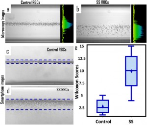
Microscopy (a-b) and smartphone (c-d) images of healthy and sickle cells distribution in the capillary; statistically significant difference between both groups distributions (e). Credit: Scientific Reports.
Sickle cell detection based on levitation in a magnetic field
The research team developed a diagnostic tool based on cell density that requires small volumes of blood and does not involve centrifugation equipment. The Sickle Cell Tester is a 3D-printed magnetic levitation platform. Red blood cells are introduced in a microcapillary where tiny magnets make them levitate. Cells are mixed with gadolinium and other chemicals that dehydrate them, causing sickle cells acquire a different density than healthy red blood cells, and a different levitation level that can be observed with the cellphone camera. Cell distribution respect to the capillary edges is analyzed with an application developed by the researchers, and the results are quickly obtained. The disease can be diagnosed without internet connection, centrifuges, microscopes or specialized staff. Patients can analyze their own blood and reduce their visits to hospitals.
In the future, the researchers plan to modify the device to make it compatible with any kind of cellphone, irrespectively of where the camera is located. They are also exploring the possibility of applying the technology to other diseases like malaria and anemia.
Source: Yale

