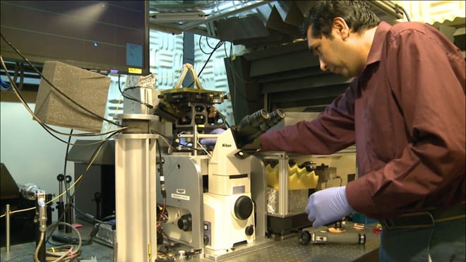
3D Force Microscopy Developed by Mizzou Researchers
A new advanced three dimensional force microscope developed by researchers from the University of Missouri now makes microscopic study of complex proteins, and their interactions possible. Until now, force microscopes were capable of delivering one dimensional results, restricting these in-depth studies.
Unlike optical microscopes, conventional force microscopes uses a needle held by a cantilever to be dragged along the surface of the sample on a slide. The compression of the needle due to the contours on the sample surface is measured by deflecting a laser beam off the cantilever, the deflection pattern of which will be recorded and results interpreted using a computer. It is necessary to prepare the samples for atomic force microscopy and these sample processing steps (crystallization or freezing) make it impossible for researchers to study these samples/specimen in its primary environment.
The new 3D force microscope developed by the researchers follows the model of a one dimensional force microscope, but with an additional laser source focused from below to measure the movement of the tip in second and third dimensions. The back scattered light from both the laser sources is collected and processed to get real-time measurements of valleys and peaks in membrane proteins along with information about the dynamic changes occurring in those structures. The new force microscope model is more flexible and can be used to study specimens in fluid conditions similar to those found inside cells to obtain more realistic results.
This new 3D force microscopy will enable researchers to study the interactions between membrane proteins on a cellular level and it will be very helpful for proteomics, drug discovery and development studies. This research is published in the journal NanoLetters.
MU Researchers Develop Advanced Three-Dimensional “Force Microscope” from MU News Bureau on Vimeo.
Source: University of Missouri


