Laser Phase Plate Becomes Reality: Biohub and Berkeley Solve Cryo-EM’s 80-Year Contrast Problem with a Laser
After more than fifteen years of being dismissed as theoretically elegant but practically impossible, the laser phase plate (LPP) has been built, installed in a working cryo-electron microscope, and shown to do what it was always supposed to do: dramatically increase image contrast for small proteins as small as 1.8Å in resolution aided by AI powered contrast.
Researchers at Biohub (the entity CZI formed last year by merging three labs and its imaging center) and UC Berkeley announced the breakthrough on June 11 across three coordinated publications — a paper in Science from Holger Müller’s Berkeley group, a theoretical framework in Nature Communications, and a not-yet-peer-reviewed Biohub preprint on bioRxiv describing a second, dual-laser design. (links below)
For structural biologists and cryo-EM core directors, this is worth paying close attention to — not because it changes anything in your facility this quarter, but because it is the first credible solution to the single biggest limitation of the technique. By Biohub’s own framing, conventional cryo-EM can resolve roughly 10% of the human proteome in purified form, and fewer than 1% of proteins in their native cellular environment. More than 90% of the proteins inside a human cell are simply too small to generate enough contrast to image clearly. The LPP is the field’s best current candidate for moving that needle, and the people behind it are careful to call it “first light through a telescope” rather than a finished instrument.
An 80-year-old Physics Problem,
Fritz Zernike described phase-contrast microscopy for light way back in 1942 and won the 1953 Nobel Prize for it: by inserting a small optical element into the light path, invisible differences in wave phase are converted into visible differences in brightness, making transparent specimens vivid. Frozen-hydrated biological samples are, to an electron beam, exactly that kind of low-contrast transparent specimen — which is why the technique has always traded away signal-to-noise on small particles.
The catch is that you cannot simply drop Zernike’s trick into an electron microscope. Any solid material placed in the electron beam gets bombarded, charges unpredictably, and ruins the image. The closest prior solution, the Volta phase plate developed in the 2010s, used a thin carbon film that exploited that charging as a feature — but it was unstable, degraded over time, and blurred high-resolution detail. That instability is precisely why the Volta plate never became routine for high-resolution single-particle work despite a decade of effort. The Science abstract is blunt about the prior art: earlier phase-plate designs were unstable and compromised high-resolution reconstruction.
The Müller–Glaeser proposal, first floated more than fifteen years ago and laid out theoretically in New Journal of Physics back in 2010, was to replace the material entirely with light. Focus a continuous-wave laser to a pinpoint at the diffraction plane, right where the electron beam passes through, and you can shift the electrons’ phase with nothing solid in the beam — nothing to contaminate, charge, or burn.
Size and Heat Made it LLP Impossible
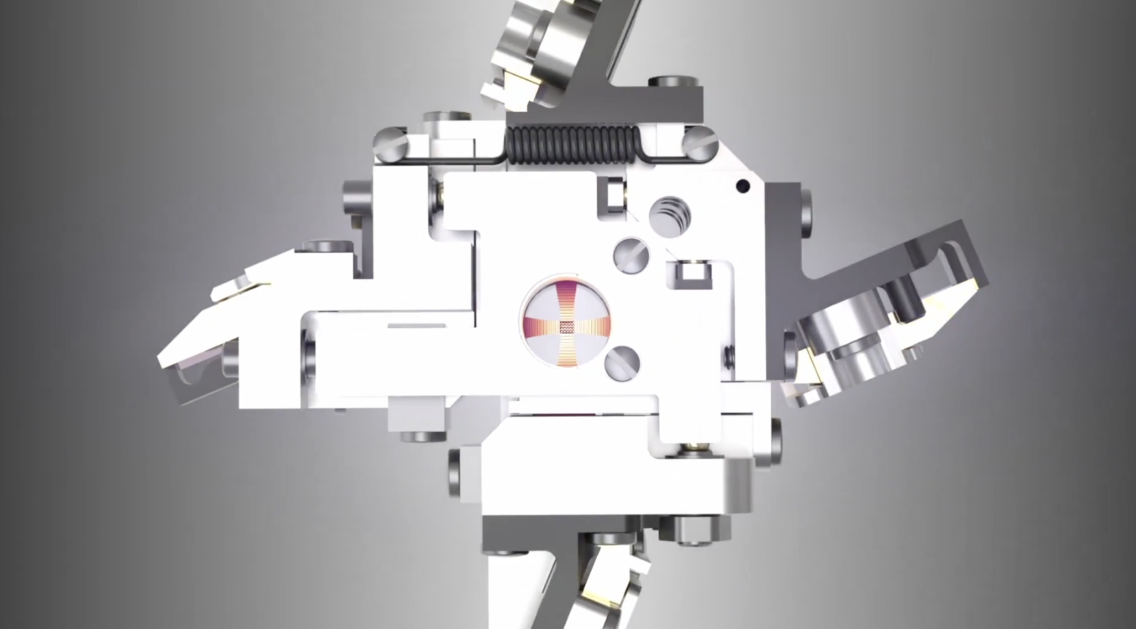
Light barely interacts with electrons, so the laser has to be extraordinarily intense — focused to a micron-scale spot, held stable for hours, and positioned to within nanometers. Müller’s solution is a Fabry–Pérot cavity: two mirrors that bounce the beam back and forth roughly 10,000 times, each pass amplifying the intensity until the focused light reaches about 100 million times the intensity of the Sun’s surface, making it the brightest continuous-wave laser of its kind in the world.
The engineering tolerances are the part that earned the “impossible” reputation. The mirrors must be polished to a surface roughness below one angstrom — roughly the diameter of a single atom — and aligned to within a thousandth of a degree. They have to stay perfectly clean: a single speck of dust will absorb the beam, burn up, and destroy the mirror. One referee on the Biohub preprint reportedly objected that nobody would ever be able to build it. Remarkably, the entire laser apparatus fits inside a device under four inches wide — “about the size of an espresso cup” — that drops into the microscope column, so from the outside an LPP-equipped instrument looks like any other cryo-EM.
The Berkeley demonstration runs on a custom Thermo Scientific Krios that Müller calls “Theia” — a “Formula 1 microscope” with extra electron optics that already out-resolves a standard Krios before the laser is even switched on. Both teams are collaborating with Thermo Fisher Scientific, the dominant cryo-EM manufacturer, which matters enormously for whether this ever leaves a handful of specialist labs.
The Dual-Laser xLPP: A More Practical Design
Arguably the more important engineering story is Biohub’s second-generation crossed laser phase plate (xLPP), which uses two laser beams in an X-shaped configuration instead of one. The dual-beam approach has two concrete advantages. First, it splits the power between two beams, so each set of mirrors runs at roughly half the intensity — meaning less stress, less catastrophic-failure risk, and tolerances that are demanding rather than borderline-impossible. Second, it suppresses ghost images — faint duplicate copies of high-contrast objects that can swamp the much fainter biological signal you actually care about.
This is where the appropriate skepticism comes in: Biohub openly states that quantifying exactly how much the xLPP reduces ghost images is still an ongoing project. The theory (the Nature Communications paper) is published; the implementation (the bioRxiv preprint) has not yet cleared peer review. The dual-laser design is the more reproducible of the two — and reproducibility, not peak performance, is the gating requirement before any of this reaches a second institution.
Not Ready for Real World Usecases
The published results are proof-of-principle on purified proteins and bacteria, not yet proteins inside intact cells. That distinction is the whole ballgame, so it is worth being precise about what has been demonstrated:
- Hemoglobin (~64 kDa) and aldolase, a standard benchmark enzyme, imaged by the Berkeley team. Hemoglobin sits near the lower frontier of what conventional cryo-EM can resolve, and with the laser on, resolution improved by up to 44% in paired laser-off/laser-on experiments. Features that were blurry sharpened into clearly defined structure.
- Apoferritin imaged on the Biohub xLPP at 1.8 Å, approaching the theoretical resolution limit of the technology — a strong signal that the laser is not trading contrast for resolution the way the Volta plate did.
- Frozen E. coli imaged with a clear contrast boost in conditions where conventional imaging struggles.
These establish that the device works, reproducibly, in a modern microscopes. They do not yet establish in-cell performance, which a few more iterations away and much harder problem.
Single-particle cryo-EM was the proving ground; cryo-electron tomography (cryo-ET) is the destination. Tomography reconstructs structures inside intact cells in 3D — not just protein shapes but how proteins interact with their neighbors — and it suffers from the contrast problem even more acutely, because tomographic samples are thicker, messier, and more crowded.
Biohub’s founding scientific director of imaging, David Agard, is direct that “the real promise is tomography.” But Bridget Carragher’s assessment is the one facility directors should internalize: cryo-ET today is “in demonstration mode, not experimentation mode, because everything is so slow.” The aspiration is to make tomography iterative — bring a biological question to the microscope, collect and analyze for a few weeks, and converge on an answer — rather than spending years grinding toward a single structure. Better contrast is also what lets you correct much of the noise inherent in tomographic data, the same way better direct-electron detectors unlocked the single-particle “resolution revolution” a decade ago. The LPP is the best candidate for that upgrade. It is not there yet.
What this means for your facility
A few practical points for core directors, structural biologists, and anyone budgeting cryo-EM capacity:
- Only two LPP-equipped microscopes exist, both in specialist hands. Whether and when Thermo Fisher commercializes the espresso-cup module — and at what price and serviceability — is the question that determines real-world impact. The seven-year, Thermo-partnered, reproducibility-first development path suggests commercialization is the explicit goal, but no timeline has been announced.
- The “valley of death” is the honest risk. Biohub itself frames the program around the gap between proof-of-concept and deployment. A device that needs sub-angstrom mirror cleanliness and nanometer stability is a serviceability and uptime question, not just a performance one. Watch for independent installations and the first results from labs outside the Biohub–Berkeley collaboration.
- The preprint is a preprint. The xLPP implementation paper has not been peer-reviewed, and the ghost-image suppression that justifies the dual-beam design is not yet quantified. Treat the dual-laser numbers as promising rather than settled.
- The data are being shared openly. Biohub is releasing its tomography data — tens of thousands of annotated tomograms — through the CryoET Data Portal, which is the kind of move that actually accelerates a field rather than a single lab. That, more than any single image, is the signal that this is being built for the community.
The bigger bet: imaging as AI training data
There is a strategic layer underneath the physics. In April, Biohub announced a five-year, $500-million Virtual Biology Initiative to generate cellular datasets for training AI models spanning molecule to organism, in health and disease. The LPP is positioned as a primary instrument for producing the high-contrast, in-cell images those models will need. In that framing, the laser phase plate isn’t only a better microscope — it’s an attempt to make the interior of the cell machine-readable at atomic resolution, feeding the same AI-for-biology convergence we’ve been tracking from the software side with Co-Scientist, ESMFold2, and the wave of structure-prediction and protein-design models.
The honest summary: What the paper does not specify is how they have dealt with the issues of heat? The setup generated 100 million times the intensity of the surface of the sun with no explanation about the solution. A single laser has literally higher flux than the flus on the sun flux. However, a device researchers spent fifteen years calling impossible now exists and demonstrably improves contrast on small purified proteins and we are optimistic. The atom-by-atom view inside intact cells — the thing that would genuinely change structural cell biology and drug discovery — is the next chapter, not this one.
Read more: Müller et al., Science (2026), DOI: 10.1126/science.aeh0665 | Crossed laser phase plate theory, Nature Communications | Biohub xLPP implementation preprint, bioRxiv | Biohub announcement | CryoET Data Portal