ScaleS: A New Technique to See Through Tissues


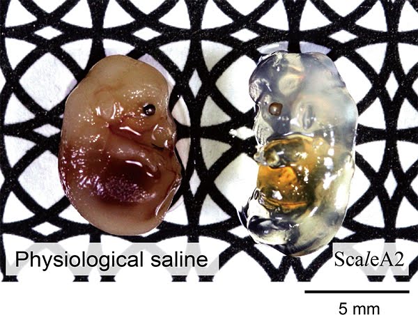
Comparison between a fetus treated with a physiological saline solution and a fetus treated with ScaleS. From: ditadifulmine.net
Japanese researchers at RIKEN Brain Research Institute have created transparent tissue. The new tissue will allow to see the brain anatomy in 3D and gain insight into neurological diseases. The study has been published in the journal Nature Neuroscience.
Traditional 2D methods can not represent the brain structure with the same accuracy as 3D methods. Optical clearing techniques, which generate “see-through” tissue, allow the use of 3D methods when combined with imaging techniques. This greater potential for the visualization of complex structures still has importants limitations, mainly the damage caused to the samples in the process of making them transparent. An example is Scale, an urea-based aqueous solution created in 2011 by Dr. Miyawaki and his colleagues. The research team spent the following years improving the solution until they found out that sorbitol combined with urea did not damage the tissues. The new solution, ScaleS, allows to obtain and store transparent brains for one year with intact structures that can bear slicing, immunolabeling, light microscopy and electron microscopy.
The authors combined ScaleS with AbScale and ChemScale, variations of the technology for immunolabeling and fluorescence, to obtain high resolution images of amyloid beta plaques in the brains of Alzheimer´s disease affected mice. They also observed human brain samples from Alzheimer´s disease patients and found out that the plaques were associated with microglia, the immune cells in the brain and spinal cord.
ScaleS and the associated techniques will allow to obtain unparallelled observation of brain structures and to detect the structural changes that are typical of the different brain diseases.
Source: genengnews

