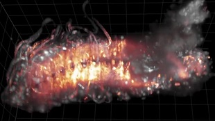
Fruit-Fly Nervous System Imaged at Nearly Single-Cell Resolution

Researchers from the Howard Hughes Medical Institute in Virginia have recorded the whole neural activity of a fruit fly for the first time. The team visualized the central nervous system (CNS) of a Drosophila melanogaster larva at almost single-neuron resolution. Genetically modified neurons emitted fluorescence when active, and an innovative microscopic technique recorded the activity. The finding was published in Nature Communications.
Functional imaging of whole CNSs has been done in larval zebrafish and C. elegans. Technical limits made it impossible to image bigger neural systems. Imaging technology could not achieve physiological timescales or higher resolution. Preparation of samples was not good enough to see whole CNS activity during behaviors. Computer methods could not interpret the activity patterns. Dr. Keller´s team addressed each of these problems to image the CNS of a larval fruit fly.
First, the team developed a multi-view imaging technique, termed high-speed simultaneous multi-view (hs-SiMView) microscopy, that improves temporal resolution in Drosophila-sized samples by 25-fold. Second, they integrated the new microscopy technique with a new sample preparation method and imaging assay for the larval CNS isolated from the rest of the body. Third, they developed a computational structure for processing and analysing the data at a rate of several terabytes per experiment. As a result, Dr. Keller and his colleagues developed the first method that records neural activity at near cellular resolution throughout the whole CNS of a higher invertebrate. the researchers characterized patterns of brain activity associated with movement, predicted functional connections between CNS regions and identified neurons with activity patterns for different motor programs.
The study opens the door for larger scale projects in the fruit-fly, but also to study the CNS of larger, more complex animals like rodents.
Source: Nature

