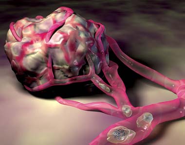
Microscopic Magnets and MRI to Detect Metastases
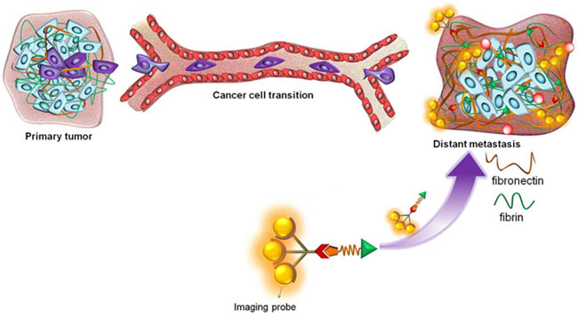
Metastases have an ECM rich in fibronectin. CWRU researchers designed a magnet-peptide chimera that binds fibrin-fibronectin complexes around metastases. This contrast agent enhances metastases identification in MRI imaging. From: Case Western Reserve University.
Scientists from Case Western Reserve University (CWRU) have presented a new technique to detect early signs of breast cancer metastases. The authors have developed a magnet-coupled peptide that binds the fibrin-fibronectin complexes surrounding metastatic areas. Magnetic resonance imaging (MRI) detects the signal emitted by the cancer-bound magnets. The finding was published in Nature Communications.
Metastasis, the spread of cancerous cells from the primary tumor to other areas of the body not directly connected, is the main cause of death in some cancer types. One third of breast cancer patients develops metastases to other organs, specially brain and lungs. Early detection of high-risk breast cancer is vital for administering timely treatment. however, current technology has difficulties to detect small high-risk breast cancer (<2 mm) and micrometastasis. MRI is widely used to visualize anatomic structures and tumors. Small molecules like chelates increase contrast of the obtained images, but they are non-specific, that is, they do not differentiate normal tissue from tumors.
A peptide linked to a micro-magnet to identify metastatic areas
Zheng-Rong Lu and his team decided to use MRI with a contrast solution that specifically detected tumors. The extracellular matrix (ECM) that surrounds cells becomes deregulated in a tumor environment. In particular, the ECM surrounding cancer cells has a much higher proportion of fibronectin (six- or seven-fold) than that of normal cells. The team decided to use the characteristic increase in fribrin-fibronectin complexes in tumors as a biomarker. They designed a short peptide that binds fibrin–fibronectin complexes in tumor ECM, and linked that peptide to a microscopic magnet that becomes magnetized during the MRI process. The team used breast cancer metastasis models from mouse and human carcinoma cells. They carried out MRI in mice bearing metastasis, 40 days after injection. This innovative contrast agent allowed to image metastases and micrometastases in distant organs such as the lung, liver, lymph node, adrenal gland and bone. Safety tests are being carried out to implement human clinical trials in the future.