Miniaturized Microscopes Allow Unprecedented Observation of Spinal Cord Cells


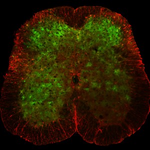
Astrocytes (green) in a spinal cord (red) react to sensation with their own chemical signals. Credit: Salk Institute.
Astrocytes, a kind nervous cells that do not conduct electrical signals, react to sensations. Thanks to tiny microscopes developed at the Salk Institute, researchers have discovered that these cells have more roles that previously thought. The new technology will help understand touch and pain phenomena in the spinal cord. The findings were published in the journal Nature Communications.
We perceive stimuli and potentially damaging environmental experiences through mechanoreceptors and nociceptors in the skin. But we still don’t know much about how this information is encoded in spinal cord cells, specially in animals behaving freely.
Axel Nimmerjahn and his team have long been working in the development of microscopes to observe the nervous system. They succeeded in the observation of brains, but the spinal cord is surrounded by multiple vertebrae, which difficults its examination. Besides, the proximity of pulsating organs (heart and lungs) difficults a stable view.
Imaging spinal cord cells in free mice
In their new study, the researchers were able to observe the activity of spinal cord cells in real time while mice where in movement. Thanks to fluorescence imaging experiments performed with miniaturized microscopes, Nimmerjahn’s team found that different stimuli activate different sets of sensory cells in the spinal cord, and that the intensity and duration of stimuli are reflected in neuronal activity. Moreover, the Salk team discovered that astrocytes, commonly considered as supporting cells, also respond to stimuli and emit chemical signals.
These new microscopes and computational processes allow unprecedented observation of spinal cord cells behavior in real time, and will improve the knowledge of nervous system development and treatment response upon injury.
Source: Salk

