A Window With a View of a Live Brain

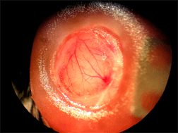
Window into a mouse’s brain. (Source: Johns Hopkins University).
A group of researchers from Johns Hopkins University have found a way to follow brain activity in real time. Through a small window in a mouse head, Rick Huganir and colleagues can watch molecular changes while the animal engage in a given observable behavior. The technique, called “brain activity mapping through imaging”, could mean a dramatic change in the way brain studies are carried out, and is already being used to study memory processes and mental illnesses.
Neuroscientists have an array of techniques to study the brain. Positron Emission Tomography (PET) and Magnetic Resonance Imaging (MRI) are techniques that obtain three-dimensional images of functional processes in the brain. Green fluorescent proteins (GFP) glow when irradiated with the proper wavelength and allow to follow neuronal responses, to track protein dynamics or identify active regions in the brain. Until recently, these techniques had to be performed either in vitro after killing the mice or in vivo but shortly after the experiment. Now, a window can be practiced on the mouse skull, replacing the bone section with a glass cover slip. Two-photon microscopy allows to visualize the fluorescent proteins, focusing the light on the desired spot, without need for slicing the brain.
Scientists are currently trying to fix a microscope in the mouse head so that they don’t need to immobilize the animal. The experiments will be more relevant as the mouse will not be constricted.
The possible applications of the new technique are astounding. There are experiments in the works for watching brain changes upon memory formation, acquisition of new experiences or astrocyte activation.
Source: Johns Hopkins