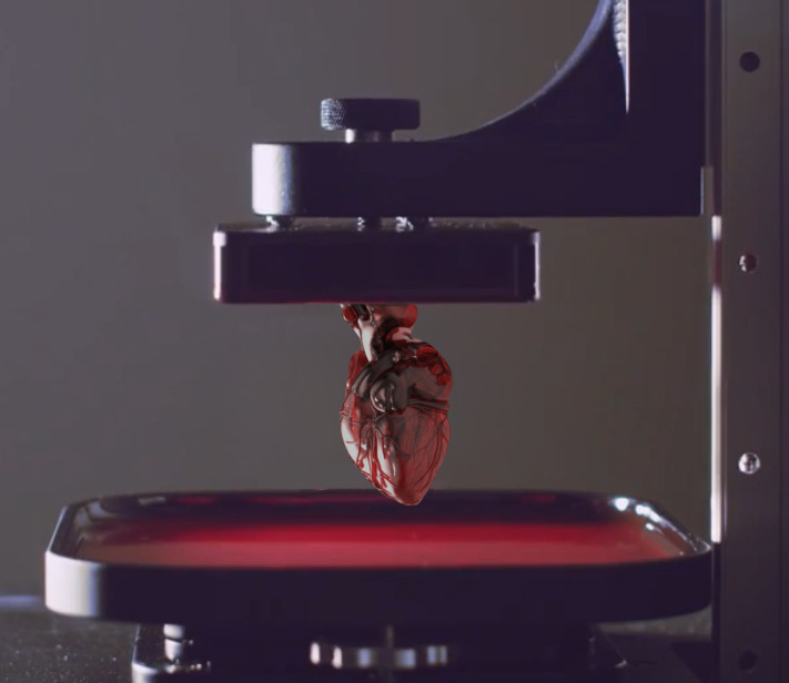
3D Bioprinting Directed by DNA “Stickers”

Scientists from UCSF have developed a new 3D bioprinting technique – DNA Programmed Assembly of Cells (DPAC) – were cells are joint together by DNA strands hanging in the outer part of the lipid bilayer. The complementarity of DNA strands allows cells to be placed like building blocks and grown in a specific fashion to develop tissues and organoids. The findings were published in the journal Nature Methods.
Cell coordination and communication is indispensable for an organ to be functional. Failure of this organizational network leads to cancer. Researchers can not study biochemical cues happening in vivo due to the complexity of isolating functions and causes of events. They could do that in a simplified organ, but in vitro tissues lack natural 3D structure. Now, DPAC allows to grow 3D organoids with the desired shape. In this technique, DNA strands with a specific sequence are tethered to the cell membrane, and they will stick like velcro to a complementary DNA hanging from other cell. As proof-of-principle, the team designed arrays of blood vessels and mammary glands. Thousands of organoids of several hundred cells each can be built in a few hours.
The DPAC technique allows researchers to specify the physical and biochemical interactions of each cell from the first stages, thus defining the development of the in vitro built tissue. Organoids could be made from a cancer patient´s cells and used as a custom drug screening platform. In vitro tissue growth could also decipher the mechanisms of organogenesis to better design complete organs in the future.
Source: UCSF

