A BioPen that Allows Surgeons to Draw Bones
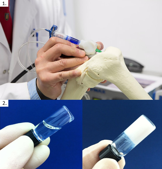
1. A handheld bio pen developed in the labs of the University of Wollongong will allow surgeons to design customised implants during surgery. 2. Injectable hydrogel scaffold undergoes rapid gelation from a soluble liquid at room temperature (left) to a stable, nonshrinking gel at body temperature (right) in a minute.[/caption]
Scientists at the University of Wollongong (UOW)-headquartered Australian Research Council Centre of Excellence for Electromaterials Science (ACES) developed the handheld BioPen to ‘draw’ new bones for orthopedic implants surgery. Meanwhile, bioengineers at the Rice University developed a temperature-sensitive hydrogel scaffold to regenerate craniofacial bone tissue,
The BioPen combines cell material within a biopolymer with another outer layer of gel material. This tri-layer ‘ink’ is solidified by an attached low powered ultra-violet light source. The cells and growth factors that are ‘drawn’ on the surgery site will then multiply and differentiate into functional bone and cartilage. However, the BioPen prototype is at its preliminary stage and optimization of cell material for use in clinical trials are to be carried out by UOW’s clinical partners at St Vincent’s Hospital, Melbourne.
At body temperature, the hydrogel developed in the Rice University lab conforms to irregular 3-D spaces, providing a platform for tissue regeneration and can be liquefied later for natural removal when sufficient bone tissue is filled at the defected site. The researchers, led by Antonios Mikos, have demonstrated the ability of the hydrogel to promote cellular attachment and proliferation in bone formation. This research was published in the journal Biomacromolecules.
Both discoveries are therefore significant in developing novel therapeutics for minimally invasive tissue regeneration.