Bruker Launches Opterra Microscope for Live Cell Imaging
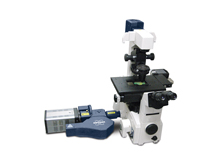

Bruker has introduced Opterra Multipoint Scanning Confocal Microscope for confocal imaging with photoactivation at the recently concluded American Society for Cell Biology Annual Meeting 2013. This new equipment with the company’s patented technology is ideal for imaging live cell preparation with minimal phototoxicity.
Opterra Multipoint Scanning Confocal Microscope uses the company’s patented swept-field imaging scanner for high speed imaging of live cells and small organisms at high resolutions while a secondary scanner performs point and area scanning for photoactivation and photoablation simultaneously. Bruker’s Opterra can be optimized for different objective lens magnifications to match with the specimen, all thanks to the seven-position pinhole/slit aperture that empowers deeper imaging of tissue in comparison with its conventional counterparts.
A whole range of photochemical techniques, utilizing various photoactivable molecules can be performed on Opterra Multipoint Scanning Confocal Microscope by coupling the photoactivation scanner with visible and multiphoton lasers. The device uses Prairie View 5.0 software for image acquisition and operating photoactivation procedures. Some of the advanced live sample studies that can be performed with Opterra includes protein localization and trafficking, vesicle and microtubule dynamics, intracellular ion imaging and nuclear structure and dynamics.
Source: Bruker