Capturing Real-time 3D Feed from Inside the Arteries


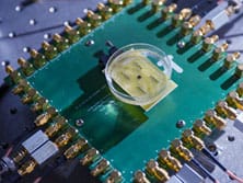
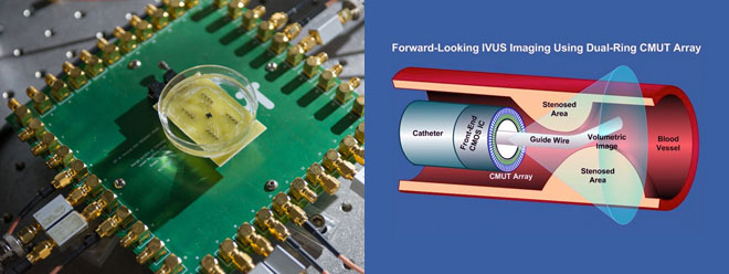
A donut-shaped silicon chip with sensing and transmitting circuitry. A thin film on top of it flutters 0.00005 of a mm, creating sound waves which are captured by an array of sensors, processed, and transmitted to an external video monitor
Scientist F. Levent Degertekin of Georgia Tech has developed a microscopic single-chip volumetric imaging system capable of capturing real-time 3D images in the arteries. This 1.5 mm donut shaped silicon chip with a 430 μm center hole for a guide wire is an integration of the CMUT (Capacitive Micromachined Ultrasonic Transducers)-on-CMOS technology which allows for miniaturization and high mechanical flexibility.
On top of the microchip is a thin film that flutters 0.00005 of a millimeter, creating sound waves captured by an a array of 100 sensors which are processed and transmitted to an external display, producing real-time volumetric ultrasound images at 60 frames per second via 13 external connections threaded through a catheter. The ability to generate such images is particularly beneficial in catheter-based clinical applications such as the diagnosis and treatment of arterial diseases. This tiny camera acts like a flashlight, allowing cardiologists to visualize blockages in occluded arteries. It is designed to consume very less power, preventing it from over-heating in a circulating blood vessel which otherwise can cause failure of the silicon elements, boiling the patient’s blood, resulting in fatalities.
Besides its current application in intravascular ultrasound (IVUS) and intracardiac echography (ICE) catheters, the potential use of this microscopic camera is vast. For instance, it can be embedded on the blades of surgical scalpels to allow surgeons to obtain a more detailed and clearer image. The development of such system is a breakthrough in medical imaging.
Source: Wired

