New AFM Based Imaging Technique Developed to Visualize Chemicals in Atomic Resolution
In one of the firsts, researchers from University of California, Berkeley have caught a single atom resolution image of a molecule in the act of rearranging its bonds. The carbon molecule from graphene, in the process of rearranging itself, has become the first molecule to be visually captured at that resolution.
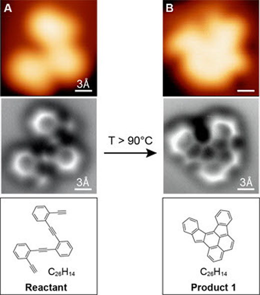
The atomic bonds, each measuring about an angstrom or more are visualized using atomic force microscopy. The images are found to be similar to the stick diagrams found in chemistry textbooks. The visualization of a molecule in atomic level represents a major breakthrough in imaging.
This new breakthrough was achieved by Felix Fischer and his team, working on graphene. They utilized the university’s powerful non-contact atomic force microscope to visualize their project involving assembly of graphene nanostructures, in order to make sure they did it right. Arranging the carbon atoms to build carbon honeycombs of repeating hexagonal patterns requires a lot of precision, as the reaction is capable of produce several different molecules.
Fischer and his team placed a ringed carbon structure on a silver plate which was heated to cause rearrangement of the molecule. And then it was cooled, trapping the resulting products. The reaction gave out an expected product as predicted along with three byproducts. The whole reaction process was visualized using advanced AFM.
This atomic resolution imaging ability can be utilized by chemists to fine tune the chemical reactions and obtain the product of interest. It will also be helpful in understanding the mechanisms involved in reactions involving various chemicals. Further reports are available in Science.
Source: UC Berkeley