New Imaging Techniques Allow Best Resolution of Living Cells of All Time
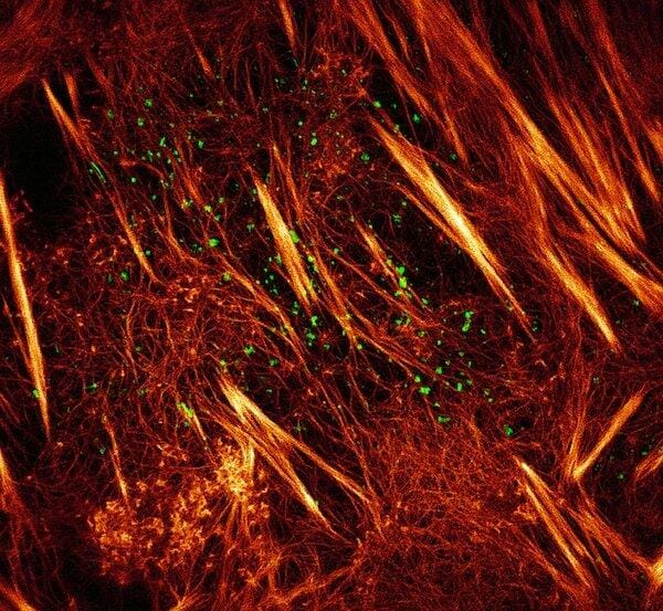

Live cell SIM of protein-pair dynamics. From: http://www.hhmi.org/news/imaging-techniques-set-new-standard-super-resolution-live-cells
Scientists at the Janelia Research Campus at Howard Hughes Medical Institute have developed new imaging methods that vastly improve resolution in structured illumination microscopy (SIM), a very advanced live cell imaging technique. The researchers have generated two new technologies based on SIM that improve the speed and non-invasiveness of image acquisition. These techniques surpass the previous resolution of 100 nm and overcome the 250 nm diffraction barrier that the bending of light dictates.The findings were published in the journal Science.
Super-resolution optical microscopy has produced the best images of subcellular structures, but could not be used in living cells. In SIM, the sample is placed under different patterns of light that are captured from different angles. The moiré images obtained are processed and converted in 3D images with twice the spatial resolution obtained with traditional means. But SIM has been long ignored because of its lower spatial resolution compared to Stimulated emission depletion (STED) and photoactivated localization microscopy (PALM).
SIM has two advantages over PALM and STED: it uses less light and it is faster. Both features prolong sample life and available acquisition time. This prompted Janelia´s researchers to try to improve its spatial resolution.
Patterned photoactivation non-linear SIM
PALM and STED act on protein samples with fotoactivable labels. All labels are photoexcited, and then most of them are turned off with another light pattern. This is necessary to produce a sharp image, as all labels turned on at the same time produce low resolution images. The problem is that most molecules are being stressed without images being taken. The researchers at Janelia found a solution: to switch on only a subset of fluorescent labels instead of all of them. this is followed by a pattern of light that deactivates the molecules. The new method, called patterned photoactivation non-linear SIM, yields 62-nanometer resolution images. The method captures 25 images for a final reconstruction in 0.33 seconds. It is very efficient, uses low intensity light and captures information from all photons emitted by the fluorescent labels. In this way, labels are conserved and scientists have more time to image cellular processes.
Patterned photoactivation non-linear SIM has already produced videos of structural proteins breaking down and reassembling, while cells move and change shape.
SIM methods can be used at no cost at Janelia’s Advanced Imaging Center by visitor researchers. However, SIM technologies will soon be accessible and affordable to any lab, as the key element is the software, not the hardware.
Source: HHMI