New Imaging Technology Reveals Chemical Composition of Tissues

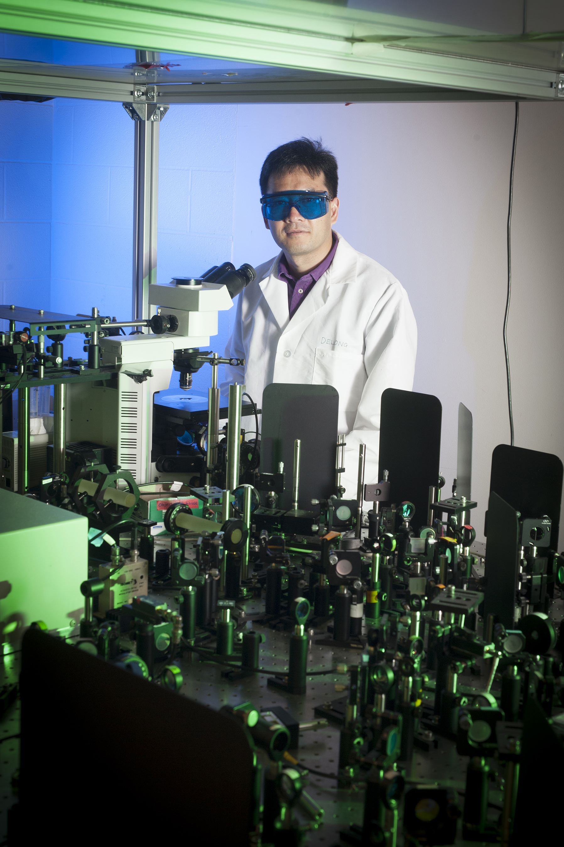
Ji-Xin Cheng and the new imaging technology for cell and tissue analysis. Credit: Purdue University/Vincent Walter.
A team of scientists from Purdue University has developed a new imaging technique that informs about the chemical composition of tissues. The new technology was possible thanks to a $1 million grant from the W.M. Keck Foundation. The study, published in the journal Science Advances, will be very useful for the improvement of medical diagnostics and cellular studies.
Current imaging technologies like tomography or magnetic resonance imaging (MRI) do not reveal the chemical composition of tissues. Optical spectroscopy techniques are not suitable for in-vivo studies: photons scatter strongly when they pass through tissues, making their detection in the spectrometer very difficult. These problems are overcome by a new technique developed by Dr. Ji-Xin Cheng and his team at Purdue.
In-vivo Raman spectroscopy
In the technique presented in this study, photons coming out of a pulsing laser are labeled with a radio frequency. After interacting with the tissue, the photons are collected by a detector. Basically, the researchers have adapted Raman spectroscopy -used for in-vitro or fixed tissue studies- to an in-vivo platform that is able to identify individual molecules in live tissues in real time. The technique does not require to label molecules, making the observations even more physiologically relevant.
The new system detected human breast cancer in two seconds, a great improvement respect to the seven days needed to do a histological examination. The technique also detected vitamin E in mice skin, showing that this method can follow topical drug delivery mechanisms.
Knowing the chemical composition of tissues will allow to detect diseases earlier than with current techniques and to conduct molecular dynamics experiments in real time. Researchers and surgeons contemplate using the technique to detect infections and to determine proper removal of cancerous tissues.
Source: Purdue