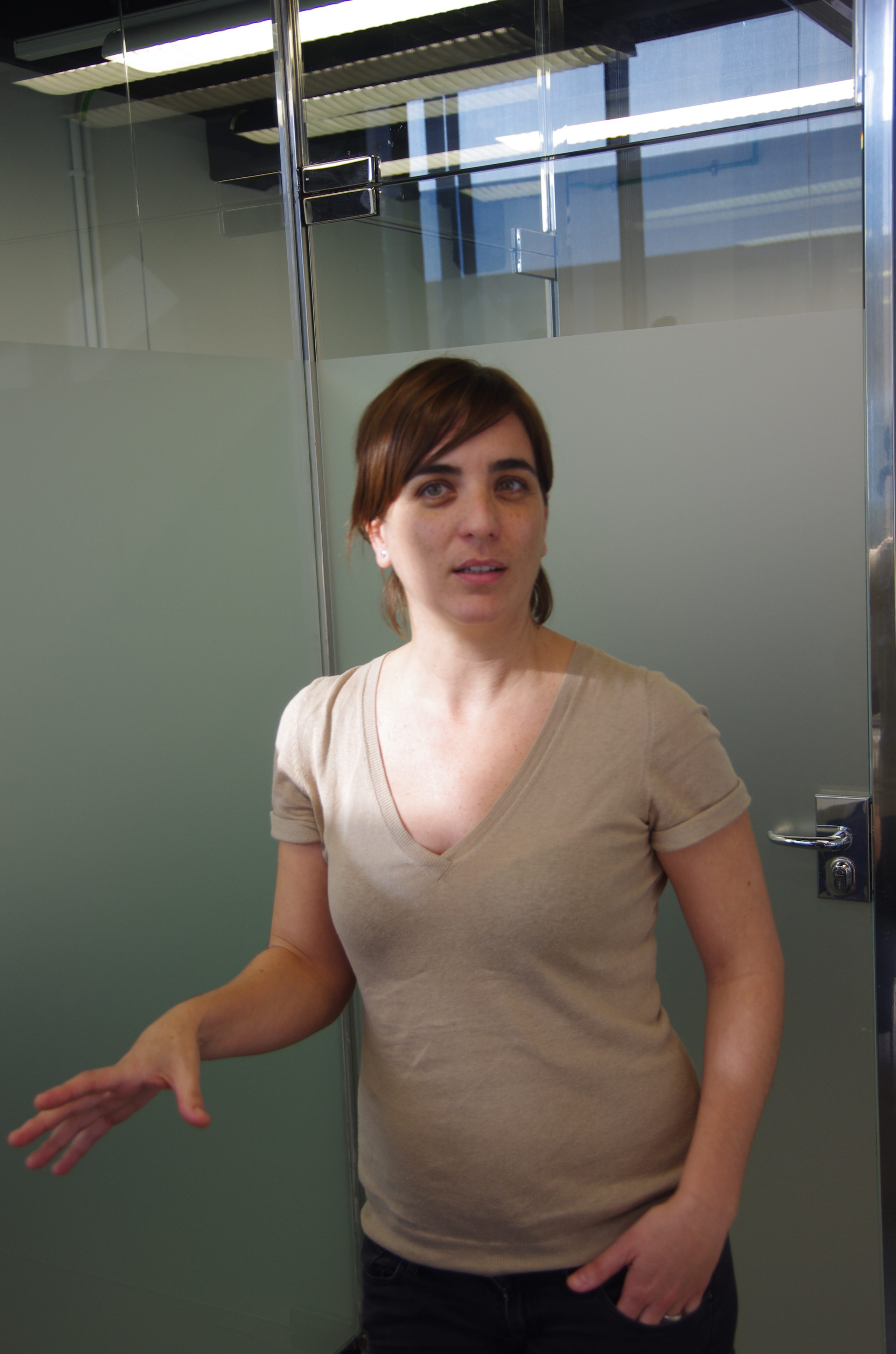
Núria Montserrat: “The Enviroment Is Fundamental for Success”
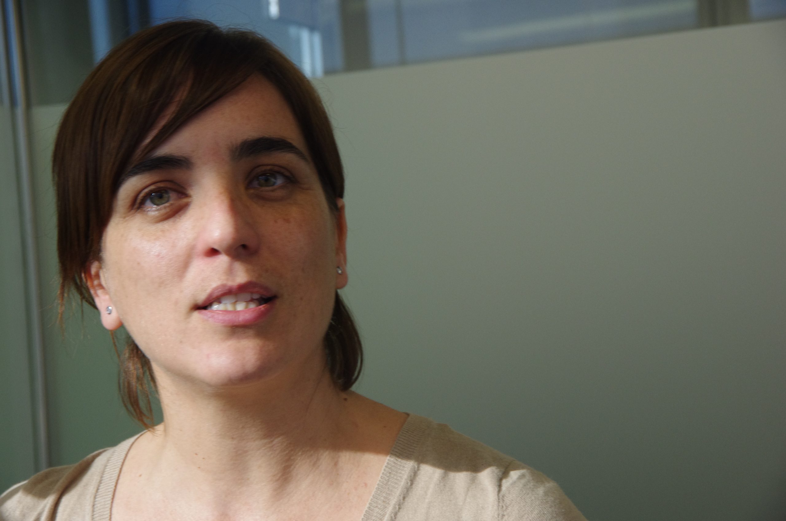
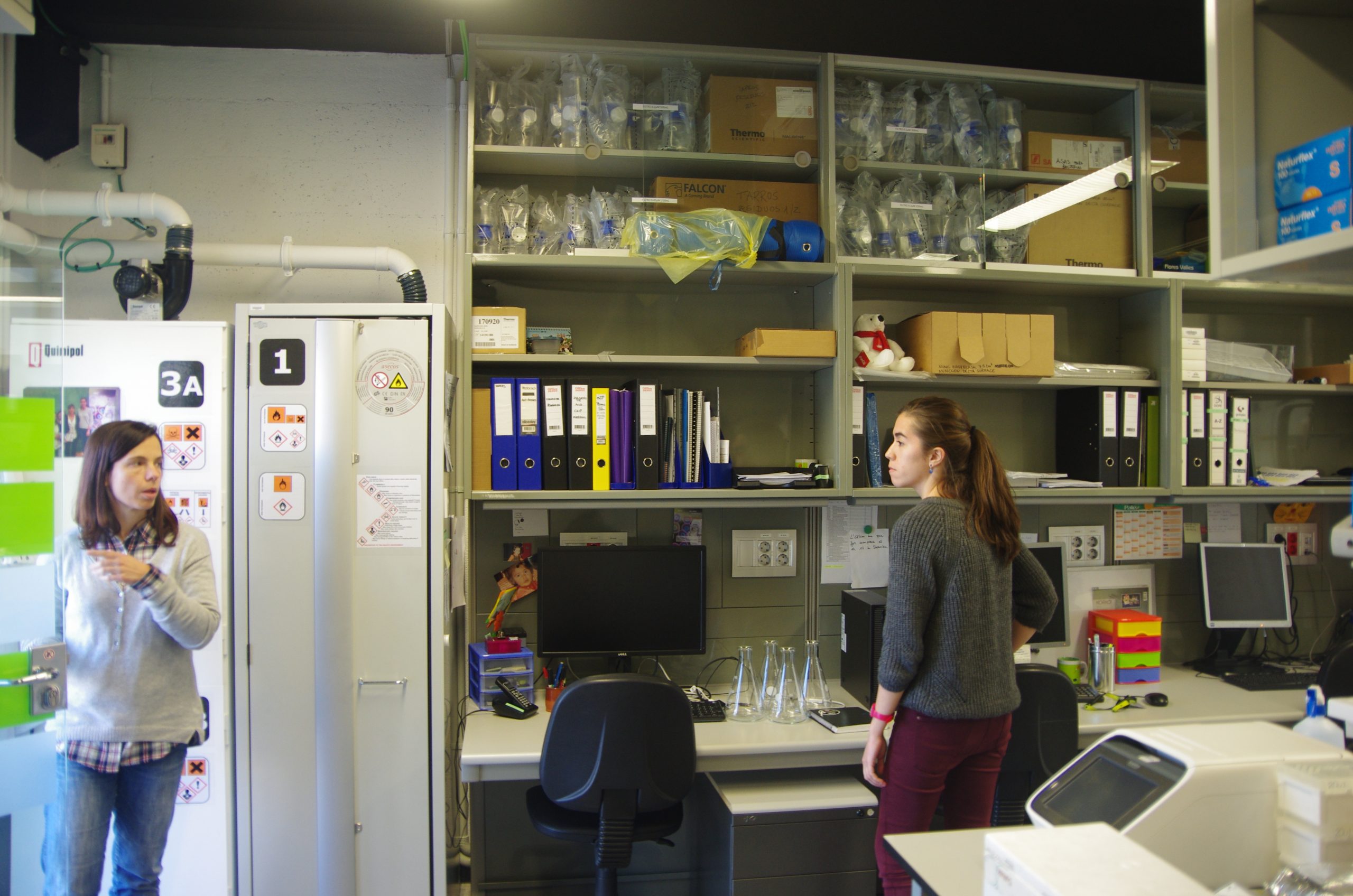
 Labcritics presents an interview with Núria Montserrrat, Junior Group Leader at the Institute for Bioengineering of Catalonia. The talented young woman shares with us her love for research, the college years, the hard -but enjoyable- PhD, the very demanding postdoc with cell-reprogramming phenomenon JC Izpisúa-Belmonte and the current days as independent researcher in the exciting fields of pluripotent stem cells and regenerative medicine. Read the transcription (in English) or watch the video (in Spanish) below.
You majored in Biological Sciences at the University of Barcelona (UB), specializing in… Biotech?
Animal Physiology and Biomedicine. Which doesn’t exist anymore.
Where those your plans, when you started studying as an undergrad?
I mean, I also really liked Biosystems, Zoology, Botany, Ecology… I always loved that other part of Biology, more traditional, more descriptive. But since I was a child I had a curiosity for these other things, more applied, more into cells… I didn’t know what I would find in the Biomedicine specialization, but more or less you could guess based on the subjects. Even if, I have to confess, I was really bad at some subjects, like biophysics. But then you take your own route and discover what you enjoy, like physiology, endocrinology… I loved those.
After your degree, you started the PhD. Was that a straightforward decision? Did you ever consider going to industry?
I was lucky… While taking a lab course in Genetics, we were crossing Drosophila mutants, I think it was the eyeless mutation… I don’t remember; it was a very basic thing. I talked to the teaching assistant and she was a 5th year undergrad. So I contacted the department in charge of the subjects I liked the most. I loved Animal Physiology and Comparative Endocrinology –from insects to mammals-. Then I became a teaching assistant myself, and I realized that the next step was the Masters. But I was lucky because I knew what I wanted to do, and that is very good to clean the “noise”, in a period of life that can be complicated, without much money, and knowing that generally PhD students struggled economically. But in my mind it was clear that I wanted to do a PhD. I could handle the instability of those years, with part-time jobs in other areas. So I was lucky. I think I was very lucky.
You started your PhD at the UB’s Department of Physiology and Immunology, under the direction of Dr. Planas.
With Dr. Planas I did the Masters. It was called Experimental Masters in Biology. It was not compulsory, it was very new, it was just starting. Not remunerated. It lasted 9 months and during that time people taking the Masters used to apply for the PhD grants that were coming out, to start the PhD afterwards. During that time I could still be in the lab, doing experiments with pro-vitelogenic ovaries from salmon and trout, a very nice and descriptive work. I eventually got a PhD grant I had applied for, and stayed in the lab. In fact, I stayed there for 8 years, with the lab classes, the Masters, the PhD and an extra year.
 The topic of the lab was… they worked with insulin and insulin-like growth factors in teleostean fish whose muscles grow indefinitely. These were basic, fundamental questions, but we had to set up a lot of things, like culturing muscle stem cells of different species and comparing the evolution of their physiological parameters. There were no openings then in the lab. The Masters was self-financed. The PhD was supported by and FPI grant. But my last year in the lab was unpaid. I mean, you know that you like what you do, and you have to make an effort to work somewhere else to cover expenses, but in that moment I knew I wanted to do that. I invested a lot of time. There were no openings, these are very hard to get positions, at the University. Some people think that if you stay long enough in the Department you end up getting a permanent position, but I think that’s not the case in that department, or in the UB, generally speaking. There might be some cases, but I know mostly people that went abroad to do a postdoc, they came back, they got a Ramon y Cajal contract, and then they got a position. People move a lot to get something like this. I don’t think it’s a matter of staying in the same lab waiting for a position. That’s my experience. The requirements are quite high.
You took a postdoctoral position at the Hospital de Sant Pau.
During the PhD I enjoyed a mobility grant and I went to the Anatomic Pathology Department of the Medical School at the University of Zurick, and later I went to the Institute of agronomy in Brittany, France. I could move around for a couple of years during my PhD. When I finished it, I went to a hospital because I wanted to experience another context, more human, more applied, therapy-focused. Human context. The fish were cool and I still read about these models, but I wanted to jump to another context.
And then you jumped to the Centre de Medicina Regenerativa de Barcelona, with Juan Carlos Izpisúa, with stays at the Salk Institute in San Diego. What did it mean to you, working with Izpisua?
It meant a boost of scientific stimulation and a way of doing things I hadn’t seen before. The fact that the director was at two places –Barcelona and La Jolla, San Diego- meant being in contact with a lot of people, getting inputs from everywhere, scientific collaborations, congresses… everything moved very fast. That change of pace, that way of doing things, is something that you don’t find easily in other environments, in this country. It’s a very different way of working. It meant opening my eyes, seeing that way of doing things and realizing that if you wanted to reach “here” you had to put effort and time. But the resources were there. It’s not about money. Izpisúa is a very proactive person and knows to delegate, so if he couldn’t help you, he knew whom you should contact to get to your goals. And that is also very hard to find: people who knows to delegate, who gives you space… It’s a very demanding environment, but in the end the benefits are for you. Those were turbulent times in the sense that I didn’t have much time for anything, but the effort totally paid off. I didn’t see it as an effort in the bad sense. Quite the opposite. A lot of good things came to me afterwards.
What was your major scientific contribution in the years you were in Izpisúa’s lab (2008-2013)?
 Mainly in the context of cellular reprogramming. We were lucky that Nobel Prize Shynia Yamanaka described cellular reprogramming in mice in 2006 and in humans in 2007, so the field was in the early stages when I joined the lab. There was one paper -by the team- in human fibroblasts and another one from an American research group. My experience was with muscle cells, satellite cells, stem cells, but I hadn’t worked with human cells. Some people on the lab had expertise in murine cells, but not many people had worked with human embryonic stem cells at the time –in a research environment. We set up protocols for reprogramming other cell types that were more permissive, like umbilical cord cells; tried other cells with a more therapeutic repercussion –e.g. mesenchymal cells-; we started correcting mutations to model diseases… And from there we jumped to tissue differentiation: we already knew how to reprogram cells, now we wanted to understand how to use reprogramming to model diseases. A lot of questions were raised, like how to efficiently derive a cardiac cell or a renal cell from an iPSC cell. Having people in the lab with background on mice stem cells was very good, it was very multidisciplinary, you could contact many people that helped in these studies. So I would say our main feats were reprogramming umbilical cord cells and obtaining mini-kidneys in culture plates.
You got a starting grant from the European Research Grant (ERC), a very prestigious, coveted and competitive grant. Did you ever imagine that you would get to where you are now?
No, not at all. In fact, I applied for the grant because Izpisúa had left the research centre and the prospects were not very good. I applied for the ERC grant taking advantage of some undeveloped ideas we were about to explore in his lab and some novel ideas that apparently nobody was working on. But I didn’t expect to get it. Just going to the interview was a major step in my career, being able to go to Brussels to defend your ideas. It’s a long process, but you have feedback, it’s nice. For a few weeks after the interview you are suffering, you don’t know what they are thinking, if they liked it. But when you get the comments from them, they even help you to set up your project. I was very lucky. I didn’t count on it, I jumped in the deep end and… many colleagues gave me advice, they helped me, they explained how the process worked… It was not premeditated, unlike everything I did up to that point. I was very lost at that point. So it was very good to count on colleagues to see where I had to put effort.
38 publications. Among them, Nature, Cell, PNAS… How is it possible to publish so much and so well in such a short time?
 I think the environment is fundamental. It’s not the money, I insist, it’s the environment in your lab, the continuous stimulation around you, the contacts you make and the desire to move forward. People might think I say so because I have the starting grant, but I didn’t have it then. You must surround yourself with a clear goal and that you can learn to dispers the chaos. You can get lost, and the environment is essential. I hear “38 publications” and I think “really?”. Those were agitated times, a lot of work, but I didn’t live through them like that. Working weekends, no vacation time, but I was happy. It wasn’t a big deal for me.
You work with hiPSCs. You take human adult cells, generally from the skin, you cultivate them in vitro, where they are de-differenciated to a pluripotent state, and from that state you can theoretically obtain any cellular type. How can you reset the epigenome to obtain this pluripotent cells?
We induce ectopic expression of transcription factors known to have a fundamental role in embryonic stem cell biology. That was described by Yamanaka long ago, in 2006. We needed lentivirus or retrovirus to express these TFs in hiPSCs. With these techniques, the TFs are integrated in the host genome. Currently, we can do iPSCs with episomal vectors: we transiently express the reprogramming factors and in 10 days we obtain iPSCs in the culture plate. Technology evolved as we were working; many companies have invested a lot of time and money in their R+D departments so that we now have culture media and matrices that allow to produce iPSCs quite systematically. We can find mutations in the original cell that prevent efficient reprogramming. This occurs, for example, when working with mutations that affect DNA repair or cell proliferation. This happens because, to reprogram a cell, we need it in a specific proliferative state, because in that moment we are demanding from the cell a reset, as you said, that implies taking the cultured cells to a well-defined state. We try to apply quite robust reprogramming systems. In some cases we find difficulties, but we have some tricks. It is known that inhibiting p53 allows better reprogramming; the presence of some cells in the medium increases reprogramming efficiency; adding feeder cells can help the reprogramming cells… We started reprogramming when there was nothing; this made us learn a lot from our mistakes. These tricks really helped us. People that started reprogramming later are used to buy the prepared “A+B” culture medium, and then find that it doesn’t work… We were lucky to start at the beginning, so we could do our troubleshooting. Some colleagues write asking how to reprogram some cells, and then I tell them to try this or that… In the same way it happens to me in fields where I am a newcomer: in theory these things are straightforward, but in practice you find problems. We not only do reprogramming and modelling in culture plate, but also differentiation. These reprogrammed cells could theoretically become any cell type from our body, at least the three cell lineages in our organism –ectoderm, mesoderm and endoderm. Today there are commercial protocols, defined media that allow you to obtain primitive endoderm, neuronal cells –not specifying very well which neurons.
 When we want to go one step beyond and model a specific disease, and a cell type that we know is affected in the pathology, which can’t be produced in vitro, we have to come up with a differentiation system ourselves, we have to read a lot of papers, understand well the development, how the intermediate mesoderm cells –which originate the kidney, gonads, adrenal gland- are formed. These cases force us to obtain knowledge either from the literature or by contacting people who works in development with that system, and start a trial and error analysis. Genome editing has opened a beautiful work in the field of stem cells. We can edit these cells’ genome, incorporate mutations that occur during the progression of a pathology, for example, or deleting mutations that we think are causing a pathology. If we have a clear phenotype and a clear question, we can reproduce the disease in a culture plate. When we speak about modelling, it’s a word that sounds very nice, but if we don’t have the question we can find ourselves with the differentiated iPSCs and not know what phenotype we should be looking for. We have to know very well the sick phenotype, so that once corrected we understand what has changed. For these reasons, it is very important to work with clinicians, people who have a different vision of the system and who can make more functional questions. Once we have corrected a mutation and we see a new phenotype, we need to be able to see that the function is recovered. There are functional tests that we don’t know how to do, or data that we don’t know to interpret. It’s important to have contact with clinicians in that area. There are 3 steps: making the iPSCs, differentiating them, and then, when we have corrected and non-corrected cells, make the questions and see if we were able to model the pathology. From here, the possibilities are huge. You can do –omics –specifically, if we have a system where we know the pathology, we can interrogate for genome changes that are influencing a given function, we can combine RNA-seq and ChIP-seq, to see new areas that we didn’t know were involved in a dysfunction, and we can find new hits to go further. All this is in vitro –we are 20 years away from the clinical stage.
When you speak about regenerating a kidney with stem cells, do you mean cell therapy, introducing cultured, functional cells in a non-functioning kidney? Could organoids also be transplanted, apart from being used to test drugs in vitro?
 This is a very interesting question… When we speak about using organoids, or cells in the kidney, we understand that we can use organoids or 3D structures to grow cells we made in the lab. We know that renal cells need a niche, a specific environment that maybe we can obtain just in bi-dimensional conditions. Being able to grow them in aggregation systems, in these 3D mini-kidneys, allows cells to grow ex-vivo. We could then take these cells and try to transplant them, if we are so lucky that the pathology only affects that specific cell type. Many times, in kidney, there are several cell types that are always affected –podocytes and tubular cells. We are probably more interested in producing these cells, auto-compatible, for transplantation. The kidney is very complex, with more than 21 cell types, with not only filtering and excretion, functions, but also hormonal ones. How can we promote endogenous regeneration, without having to implant cells, how can we trigger auto-regeneration in the kidney? There are resident stem cells in the kidney, with a quite good turnover, and there is a lot of discussion about them, but we know that they are involved in renal pathology in aging. They act like myosatellite cells. With those cells we could reestablish parts of the nephron. We can’t make a new nephron. Those cells can’t make new nephrons like they do in zebrafish or in insects, that form a new structure when the kidney is damaged. In mammals, maybe due to evolution, the organ complexity required more specialization and regenerative capacity was lost. Resident stem cells replace parts of the structure, but not the whole structure. We are trying to understand the regenerative signals that occur in other organisms and even in mammals during development to be able to stimulate those responses in the adult kidney. We know that some hits, some candidates that seem to help to regenerate the kidney endogenously by overexpression or downregulation. There is another problem: cell therapy is very complex, all the regulation steps, understanding the cell as a drug, the legal part of how to take the iPSCs to the clinic… There are many groups trying to do that. Prof. Yamanaka did a study in retina, he did iPSCs from patients with age-associated macular degeneration, and they found that there were problems with the first patients. It’s not going to be easy to go to the clinical step. We use iPSCs as a tool to understand development, pathologies in the culture plate, and to understand which signals promote regeneration, which would spare the use of cells; we would just apply endogenous molecules or compounds that activate endogenous pathways. It is very risky in the renal field; many people were doing kidney research before me, they have more background, and they are taking a different approach, trying to promote endogenous regeneration in mammals.
How do you generate a 3D organoid? Do you use any polymer, 3D-printers?
 We aggregate human cells using murine cells, with murine embryonic kidney cells. There is a time window in the development of these cells where we can disaggregate and reaggregate with our pre-differentiated hiPSCs. We put them in organotypical growth conditions, which is a complex system, cells then reaggregate and we don’t have to do much because that’s what they know to do. If you take cells out of the kidney and put in vitro, they will reaggregate and try to make the structure they had inside the animal. We use these reaggregation systems mixing human cells; there is a percentage we can mix and aggregate, like 90% of murine cells with 10% human cells, and we can keep this in culture. Another way of doing it, recently published by Melissa Little’s group, is to make 3D structures using collagen, or low-attachment systems, allowing cells to reaggregate during differentiation. They haven’t used any other species’ cells for reaggregation. In 18 days, after culturing cells in bi-dimensional systems, they disaggregate them and the cells reaggregate, and with the presence of cytokines in the culture medium they obtain nephrons spontaneously, let’s say. We haven’t reached this point, because we think it’s very interesting to understand which signals give rise to aggregation, how can we mimetize them. We don’t care much about co-culturing two cell types from different species. If the tag out cells with a fluorescent protein, a reporter, after growing and aggregating them we can recover and analyze them.