RNA Response to Drugs Visualized in Living Mice Brains

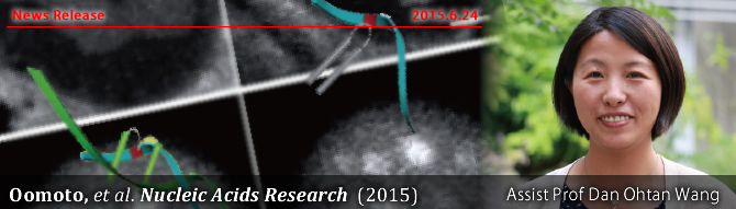
A Japanese research group has observed RNA dynamics in vivo for the first time. The researchers used a new method for high-resolution fluorescence RNA imaging, ECHO-liveFISH. This technique enabled the observation of nuclear RNA foci in mice and chicks by RNA fluorescent labeling. This finding will help identify hot-spots of gene expression and compare expression of healthy and sick tissues in living beings. The work was published in Nucleic Acids Research.
RNA molecules are translated into proteins, the molecular machinery that makes cells work. Differences in RNA distribution and density inside the cell can thus account for very different biological outcomes. It is not well known how RNA is distributed and the reasons for that. Studying RNA dynamics would help understand gene expression and engineer a given locus if necessary. Efforts done in the past to visualize RNA were hampered by the lack of a good in vivo RNA imaging technique. But Professor Dan Ohtan Wang from Kyoto University’s Institute for Integrated Cell-Material Sciences (iCeMS) and his team designed a new technique based on fluorescent probes.
Drug screening in a physiological setting
Ohtan and colleagues designed fluorescent, non-toxic probes that bound to RNA inside the neurons of live mice. They could track the RNA movements inside the cell and make quantitative analyses by measuring the fluorescence emission level. The study also proved that RNA response to drugs is different in vivo than in cultured cells. Drug-screenings will therefore be more physiologically relevant thanks to this new technique.
Source: iCeMS Kyoto University