Spinning Disk Statistical Imaging (SDSI) System: The Improved Super-Resolution Microscopy
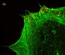
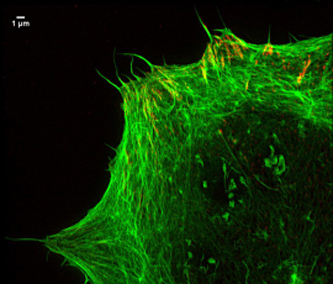
Focal adhesions and actin
A team of researchers (Dr. Neveen Hosny, a bioengineer from Prof. Martin Knight, School of Engineering and Materials Science; Dr. Ann Wheeler, Head of Imaging at Queen Mary’s Blizard Institute) from the Queen Mary University of London have developed a Spinning Disk Statistical Imaging (SDSI) system that offers super-resolution imaging of a range of cellular structures. This system is an improved technique addressing the limitations of super-resolution microscopy, which can only visualize structures at the bottom of the cell thereby unable to locate the nucleus, bacterial and viral infections occurring in the centre and anywhere else in the cell.
The SDSI system is a combination of a spinning disk confocal microscope and Photoactivation Light-Microscopy (PALM)/Stochastic Optical Reconstruction Microscopy (STORM) biological probes, providing flexibility and simplicity. This newly developed system allows fast, reliable and more detailed imaging of structures less than 80 nm anywhere in the cell. As indicated by the name of the microscope, high speed imaging was achieved by using a disk with an array of tiny holes that remove out of focus light.
The researchers are looking forward to using this system in high-content screening, imaging of multiple epitopes and live cell imaging which can be applied in the study of stem cell behaviour to better understand arthritis and the development of nanomedicine as well as cancer invasion.
Source: Queen Mary University of London