New 3D Bioprinting Method Obtains Heart Model

Credit: Carnegie Mellon University.
Researchers from Carnegie Mellon University have printed 3D models of hearts, bones, arteries and brains using soft biological materials. The models were based on 3D high-resolution images of real organs. The new technique solves the problems in using gels for bioprinting. The study has been published in the journal Science Advances.
3D printing has progressed enormously in the past years. The traditional method uses hard substances like plastic or metal that are deposited layer by layer. Lower layers support the upper ones, which has so far hindered the use of softer printing materials, especially for biomedical applications, where collagen or fibrin are crucial to engineer tissues. The gelatinous materials collapse when new layers are placed on the structure.
Freeform Reversible Embedding of Suspended Hydrogels
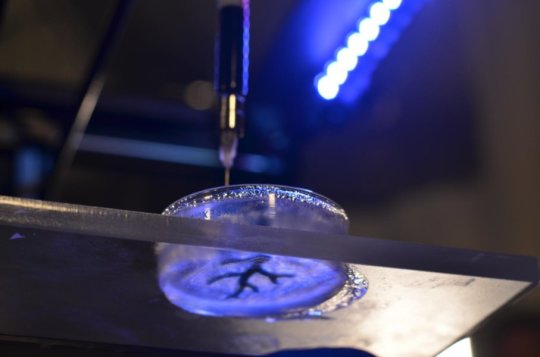
The researchers developed a new technique that enables them to obtain 3d biostructures from gelatinous materials. The team obtained 3D images of several organs at high resolution by methods like tomography and magnetic resonance; then, they used these images to construct the 3D models at 200um resolution by their method, Freeform Reversible Embedding of Suspended Hydrogels (FRESH). It consists of inserting the protein and polysaccharide hydrogels that will constitute the organ into a secondary hydrogel structure, a temporary, thermoreversible support. One gel is printed inside another gel, layer by layer. When the structure is finished, the outer case is removed by heating to body temperature, that causes the supporting gel to melt while not affecting the biomolecules that constitute the bioprinted organ model.
Dr. Feinberg´s group has implemented their technique in the consumer-grade $1000 3D-printers, far cheaper than the $100000 that usually costs a top of the line bioprinter. It does not require specialized staff, and hardware and software are open-source, which allows to modify the printing settings. The research team has released the 3D printer designs under a free-use license.
In the future, the researchers plan to insert real cardiomyocytes into the bioprinted structure, which would work as a platform for the formation of a functioning heart.
Source: CMU