Shimadzu Introduces iMScope Imaging Mass Microscope
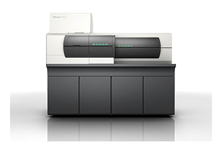

Shimadzu Corporation, the leaders in analytical instruments has recently launched iMScope, a novel imaging mass microscope. The new device has both optical microscope and a hybrid ion TOF mass spectrometer for observing high resolution morphological images and for identifying and visualizing the distribution of molecules in the specimen sample respectively.
Shimadzu’s iMScope offers a mass spectroscopy resolution 5μm to enable visualization of molecular distribution in sub cellular levels. iMScope employs the company’s proprietary ultra focusing laser optics along with 3D automated sample transfer stage. Living cells and tissue samples can be analyzed by atmosphere MALDI. The mass spectrometry images are superimposed with morphological images from optical microscope to reveal the molecular distribution pattern in the specimen sample and its relation to biological functions or morphological changes.
Traditional mass spectrometric analysis requires extraction and homogenization of samples for pre-treatment, which leads to loss of positional information of molecules in the sample and makes it know where a particular molecule was located and the nature of molecules populating the region of interest. It is an issue faced by researchers working on disease biomarker research and biological function studies. This issue is addressed by iMScope.
In iMScope, the sectioned sample is first irradiated by laser at different sample points and the ionized molecules in these regions are analyzed by the mass spectrometer. The two dimensional distribution of specific molecules are then easily visualized by combining the positional information of each mass spectrum and the relative amounts of specific ions in the spectrum to produce an accurate map of the distribution pattern of these molecules.
Source: Shimadzu