Super-Resolution Microscopy to Diagnose Rare Platelet Disorder
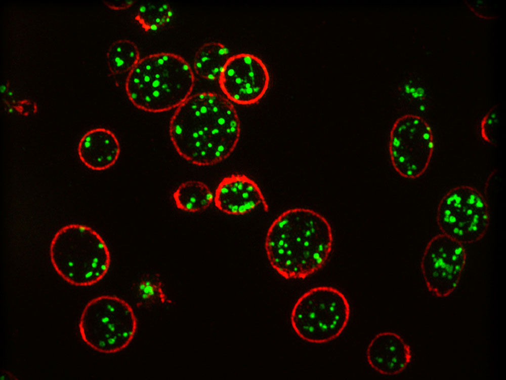

SIM image of platelets. Credit: Professor Dan Cutler, UCL.
A team of scientists led by a research group at University College London have developed a super-resolution microscopy method to diagnose a rare bleeding disease. The new method is more accessible and allows a more accurate diagnosis of platelet disorders. The results were published in the Journal of Thrombosis and Haemostasis.
Platelets are blood components that adhere to damaged blood vessels and stop bleeding. After the first response to maintain homeostasis, the platelets activate a coagulation cascade and release several kinds of granules, loaded with coagulation factors, at the site of injury. There are many types of platelet disorders, but a diagnostic technique that can differentiate them is lacking, which difficults the application of the proper treatment. Electron microscopy is the technique of choice: it has resolutive power to observe the granules. However, it is expensive, it can’t be found in every clinical setting, it needs fresh samples and it often leads to wrong diagnoses.
Structured Illumination Microscopy
The researchers decided to try to diagnose platelet disorders with Structured Illumination Microscopy (SIM), a type of super-resolution microscopy. In SIM, samples don’t need to be analized right after being collected. As a proof of concept, they tested samples from healthy people and from individuals with the Hermansky-Pudlak syndrome, a blood disorder that affects 1 in 500000. They tagged platelet granules with the marker protein CD63 and observed them with a custom-built system that automatically counted the number of granules per platelet. With the automated system, which ensures unbiased and faster analysis, the researchers correctly diagnosed the disease with 99% confidence, compared to 25% of correct initial diagnoses with previous techniques. The system detected that sick people had one third as many granules as healthy people.
The researchers expect that their study will inspire others to use super-resolution microscopy to analyze and diagnose other blood disorders, which allows to test multiple parameters by tagging the appropriate molecule.
Source: UCL