Tumour-on-a-chip Device to Revolutionize Personalized Treatment for Cancer
Researchers from University of Toronto’s Institute of Biomaterials and Biomedical Engineering (IBBME) have developed a new tumour-on-a-chip microfluidics screening platform for predicting the behaviour of nanoparticles in the living body. This new device, with a potential to revolutionize personalized cancer treatment is designed to expedite the study of behaviour and effects of nanoparticles on tumour tissue in living body by using microfluidics technology.
Finding new effective treatments for cancer is one of the major areas of research in life sciences. A significant part of this research is focused on developing targeted drug delivery systems. Nanoparticles are the drug delivery vehicles of choice because of their size and reach inside the human body. These nanoparticles can be easily used to mark the tumours or even deliver chemotherapy agents directly to cancerous cells with increased potency and reduced side effects. Generally, the outer surfaces of these nanoparticles are treated to make them stick to the surface of the tumour cells, which works fine in the in-vitro conditions. But while in-vivo, these treated nanoparticles cling on to the surface of these tumour cells, without penetrating into the tumour to deliver the drugs or treat the affected tissue.
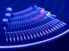
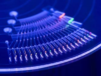
Until now, the only ways to study these nanoparticles have been either on petri dishes or by injecting them into mice for live testing. One of the drawbacks of live testing is that the scientists can only observe the final destination of nanoparticles inside the body, not the path taken by them to travel from the injection site to target site or their behaviour during that period. The tumour-on-a-chip designed by the researchers is a microfluidic device in which live spheroid tissues grown from biopsies, mimicking cancerous tumours are placed inside an inch long chamber with saline solution constantly flowing through it, to reproduce the conditions found in tumours. By injecting fluorescent nanoparticles into the chamber, researchers can find the number of nanoparticles penetrating the tissue, their accumulation pattern and the effect of liquid speed on the movement of nanoparticles.
The experiments conducted on the tumour-on-a-chip devices predicted the behaviour of nanoparticles in larger live models within an hour as compared to weeks, taken by conventional live testing method in mice. The efficacy of different nanoparticles for spheroid tissues grown from patient’s biopsy can be tested using this device to select the suitable nanoparticle for treatment, making personalized treatment easier. This technology has a huge potential to simplify cancer screening and treatment.
Source: University of Toronto