
A Non-Toxic DNA Stain for Super Resolution Microscopy
Scientists from the Échole Polytechnique Fédérale de Laussane (EFPL) have created a new DNA stain that can be used in live cells. The far-red DNA stain, called...
Basic Research
Optical, electron and emerging microscopy methods, and the advances pushing resolution and speed.
35 articles
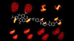
Scientists from the Échole Polytechnique Fédérale de Laussane (EFPL) have created a new DNA stain that can be used in live cells. The far-red DNA stain, called...
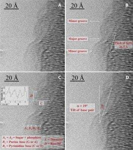
Researchers at King Abdullah University of Science and Technology (KAUST) in Saudi Arabia have developed a technique that allowed them to visualize directly the DNA helix...
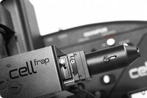
Olympus presents the cellFRAP, a live cell imaging platform for the in-vivo analysis of dynamic processes by accurate and flexible photomanipulation techniques. cellFRAP´s main feature is...

Scientists at the Janelia Research Campus at Howard Hughes Medical Institute have developed new imaging methods that vastly improve resolution in structured illumination microscopy (SIM), a very advanced live cell...
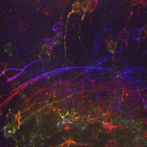
Bruker has announced the release of the Opterra II™ Multipoint Scanning Confocal Microscope. The Opterra II™ is a high speed, quantitative live-cell miscroscope characterized by low photo-toxicity and...
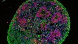
Based on studies on lab-grown cerebral organoids, autism could be linked to an excess of inhibitory neurons. Brain models derived from the skin cells of...
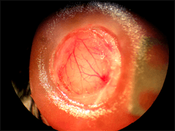
A group of researchers from Johns Hopkins University have found a way to follow brain activity in real time. Through a small window in a...
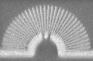
Scientists at the University of Buffalo are pushing the limits of microscopy with their new hyperlens. Currently in prototype stage, this new hyperlens is developed...